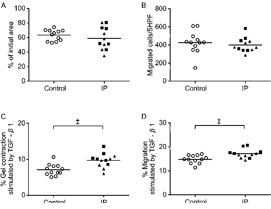
Fig. 1. Functional assays with control and lung fibrotic (interstitial pneumonia, IP) fibroblasts. (A) Contraction of three-dimensional collagen gels. Fibroblasts were cultured and cast into three-dimensional collagen gels that were maintained in suspension, with gel size measured daily. The vertical axis shows the gel size after 3 days of contraction, expressed as a percentage of the original size. (B) Chemotaxis. Fibroblasts were grown in a monolayer culture, trypsinized, and their chemotactic activity towards fibronectin (20 µg/ml) was assessed. The vertical axis shows the number of migrated cells per five high-power fields (5HPF). (C) Effect of TGF-β1 on gel contraction. The vertical axis shows the ratio of the size of TGF-β1-stimulated gels to that of untreated gels on day 3. (D) Effect of TGF-β1 on chemotaxis. The vertical axis shows the ratio of the number of migrated cells in response to TGF-β1 treatment to that of migrated cells without treatment. Responses of fibroblasts from control and IP subjects to TGF-β1 in the gel contraction and chemotaxis assays were assessed as described in the Materials and Methods. Closed triangles and squares indicate lung fibrosis subjects with histological nonspecific interstitial pneumonia and usual interstitial pneumonia patterns, respectively. Each symbol represents one patient. The values represent the mean ± SEM of at least three independent experiments. ‡P<0.005.